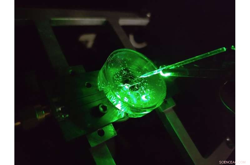
Un prototype d'un microscope d'imagerie à tension de diamant construit par des physiciens de l'Université de Melbourne. Une minuscule électrode est suspendue au-dessus de la puce de diamant pour tester les performances de l'appareil. Un laser vert éclairé par le bas fournit une excitation de fluorescence à la puce. Crédit :Auteur fourni, Université de Melbourne
Le cerveau est sans doute l'une des structures les plus complexes de l'univers connu.
Les progrès continus dans notre compréhension du cerveau et notre capacité à traiter efficacement une multitude de maladies neurologiques reposent sur l'exploration des micro-circuits neuronaux du cerveau avec des détails de plus en plus détaillés.
Une classe de méthodes pour étudier les circuits neuronaux est appelée imagerie de tension. Ces techniques nous permettent de voir la tension générée par les neurones de déclenchement de notre cerveau, nous indiquant comment les réseaux de neurones se développent, fonctionnent et changent au fil du temps.
Aujourd'hui, l'imagerie de tension des neurones cultivés est réalisée à l'aide de réseaux denses d'électrodes sur lesquelles les cellules sont cultivées (ou cultivées), ou en appliquant des colorants électroluminescents qui répondent optiquement aux changements de tension à la surface de la cellule.
Mais le niveau de détail que nous pouvons voir en utilisant ces techniques est limité.
Les plus petites électrodes ne peuvent pas distinguer de manière fiable les neurones individuels, environ 20 millionièmes de mètre de diamètre, sans parler du réseau dense de connexions à l'échelle nanométrique qui se forme entre eux, et aucune avancée technologique significative n'a été réalisée dans ce domaine depuis plus de deux décennies.
De plus, chaque électrode nécessite sa propre connexion filaire et son propre amplificateur, ce qui limite considérablement le nombre d'électrodes pouvant être mesurées simultanément.
Les colorants peuvent surmonter ces limites en imaginant la tension sans fil sous forme de lumière, ce qui signifie que l'électronique complexe peut être située loin des cellules d'une caméra.
Le résultat est une haute résolution sur de grandes surfaces, capable de distinguer chaque neurone individuel dans un grand réseau. Mais il y a aussi des limites ici, les réponses en tension des colorants de pointe sont lentes et instables.
Nos recherches récentes publiées dans Nature Photonics , explore un nouveau type de plate-forme d'imagerie de tension haute vitesse, haute résolution et évolutive créée dans le but de surmonter ces limitations :un microscope d'imagerie de tension en diamant.
Développé par une équipe de physiciens de l'université de Melbourne et de l'université RMIT, l'appareil utilise un capteur à base de diamant qui convertit les signaux de tension à sa surface directement en signaux optiques, ce qui signifie que nous pouvons voir l'activité électrique en temps réel.
The conversion uses the properties of an atom-scale defect in the diamond's crystal structure known as the nitrogen-vacancy (NV).
NV defects can be engineered by bombarding the diamond with a nitrogen ion beam using a special type of particle accelerator. The fabrication of the sensor begins with using this process to create a high-density, ultra-thin layer of NV defects close to the diamond's surface.
You can think of each NV defect as a bucket that holds up to two electrons. When this bucket is empty, the NV defect is dark. With one electron, the NV defect emits orange light when illuminated by a laser—this property is known as fluorescence. With two electrons, the color of the fluorescence becomes red.
A previously discovered property of NV defects is that the number of electrons they hold—and the resulting fluorescence—can be controlled with a voltage. Unlike dyes, the voltage response of an NV defect is very fast and stable.
Our research aims to overcome the challenge of making this effect sensitive enough to image neuronal activity.
On the diamond's surface, the crystal structure ends with a layer one atom thick, made up of hydrogen and oxygen atoms. The NV defects closest to the surface are the most sensitive to changes in voltage outside the diamond, but they are also highly sensitive to the atomic makeup of the surface layer.
Too much hydrogen and the NVs are so dark that the optical signals we are looking for cannot be seen. Too little hydrogen and the NVs are so bright that the small signals we are after are completely washed out.
So, there's a "Goldilocks' zone" for voltage imaging, where the surface has just the right amount of hydrogen.
To reach this zone, our team developed an electrochemical method for removing hydrogen in a controlled way. By doing this, we've managed to achieve voltage sensitivities two orders of magnitude better than what has been previously reported.
We tested our sensor in salty water using a microscopic wire 10-times thinner than a human hair. By applying a current, the wire can produce a small cloud of charge in the water above the diamond. The formation and subsequent diffusion of this charge cloud produces small voltages at the diamond surface.
By capturing these voltages through a high-speed recording of the NV fluorescence, we can determine the speed, sensitivity and resolution of our diamond imaging chip.
We were able to further boost sensitivity by patterning the diamond's surface into 'nanopillars'—conical structures with the NV centers embedded in their tips. These pillars funnel the light emitted by the NVs towards the camera, dramatically increasing the amount of signal we can collect.
With the development of the diamond voltage imaging microscope for detecting neuronal activity, the next step is the recording of activity from cultured neurons in vitro—these are experiments on cells grown outside their normal biological context, otherwise known as test-tube or petri-dish experiments.
What differentiates this technology from existing state-of-the-art in vitro techniques is the combination of high spatial resolution (on the order of a millionth of a meter or less), large spatial scale (a few millimeters in each direction—which for a network of neurons in mammals is quite vast), and complete stability over time.
No other existing system can simultaneously offer these three qualities, and it's this combination that will allow our made-in-Melbourne technology to make a valuable contribution to the work of neuroscientists and neuropharmacologists globally.
Our system will aid these researchers in pursuing both fundamental knowledge and the next generation of treatments for neurological and neurodegenerative diseases. New method enables long-lasting imaging of rapid brain activity in individual cells deep in the cortex