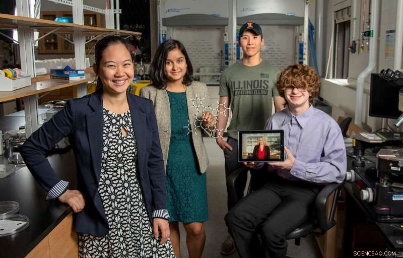
Pinshane Huang, à l'extrême gauche, rejoint, Priti Kharel, deuxième à gauche, Justin Bae et Patrick Carmichael, à l'extrême droite, au Laboratoire de recherche sur les matériaux. Sur la tablette, on voit Blanka Janicek. Crédit :Heather Coit/The Grainger College of Engineering
Un effort de recherche de l'Université de l'Illinois à Urbana-Champaign dirigé par Pinshane Huang accélère les techniques d'imagerie pour visualiser clairement les structures de petites molécules, un processus autrefois considéré comme impossible. Leur découverte libère un potentiel infini dans l'amélioration des applications de la vie quotidienne, des plastiques aux produits pharmaceutiques.
Le professeur agrégé du Département de science et génie des matériaux s'est associé aux co-auteurs principaux Blanka Janicek, ancienne élève de 21 ans et post-doctorante au Lawrence Berkeley National Laboratory à Berkeley, en Californie, et Priti Kharel, étudiante diplômée du Département de chimie, prouver la méthodologie qui permet aux chercheurs de visualiser de petites structures moléculaires et d'accélérer les techniques d'imagerie actuelles.
Les co-auteurs supplémentaires incluent l'étudiant diplômé Sang hyun Bae et les étudiants de premier cycle Patrick Carmichael et Amanda Loutris. Leurs recherches évaluées par des pairs ont récemment été publiées dans Nano Letters .
Les efforts de l'équipe exposent la structure atomique de la molécule, permettant aux chercheurs de comprendre comment elle réagit, d'apprendre ses processus chimiques et de voir comment synthétiser ses composés chimiques.
"La structure d'une molécule est tellement fondamentale pour sa fonction", a déclaré Huang. "Ce que nous avons fait dans notre travail, c'est de permettre de voir directement cette structure."
La capacité de voir la structure d'une petite molécule est vitale. Kharel partage à quel point il est vital en donnant l'exemple d'un médicament connu sous le nom de thalidomide.
Découverte dans les années 60, la thalidomide était prescrite aux femmes enceintes pour traiter les nausées matinales et s'est avérée plus tard causer de graves malformations congénitales ou, dans certains cas, même la mort.
Qu'est ce qui ne s'est pas bien passé? Le médicament avait des structures moléculaires mixtes, l'une responsable du traitement des nausées matinales et l'autre causant malheureusement des effets néfastes dévastateurs sur le fœtus.
Le besoin d'une science proactive et non réactive a poussé Huang et ses étudiants à poursuivre cet effort de recherche qui a commencé à l'origine par pure curiosité.
"Il est tellement crucial de déterminer avec précision les structures de ces molécules", a déclaré Kharel.
En règle générale, les structures moléculaires sont déterminées avec des techniques indirectes, une approche longue et difficile qui utilise la résonance magnétique nucléaire ou la diffraction des rayons X. Pire encore, les méthodes indirectes peuvent produire des structures incorrectes qui donnent aux scientifiques une mauvaise compréhension de la composition d'une molécule pendant des décennies. The ambiguity surrounding small molecules' structures could be eliminated by using direct imaging methods.
In the last decade, Huang has seen significant advancements in cryogenic electron microscopy technology, where biologists freeze the large molecules to capture high-quality images of their structures.
"The question that I had was:What's keeping them from doing that same thing for small molecules?" Huang said. "If we could do that, you might be able to solve the structure (and) figure out how to synthesize a natural compound that a plant or animal makes. This could turn out to be really important, like a great disease-fighter," Huang said.
The challenge is that small molecules are often 100 or even 1,000 times smaller than large molecules, making their structures difficult to detect.
Determined, Huang's students began using existing large molecule methodology as a starting point for developing imaging techniques to make the small molecules' structures appear.
Unlike large molecules, the imaging signals from small molecules are easily overwhelmed by their surroundings. Instead of using ice, which typically serves as a layer of protection from the harsh environment of the electron microscope, the team devised another plan for keeping the small molecules' structures intact.
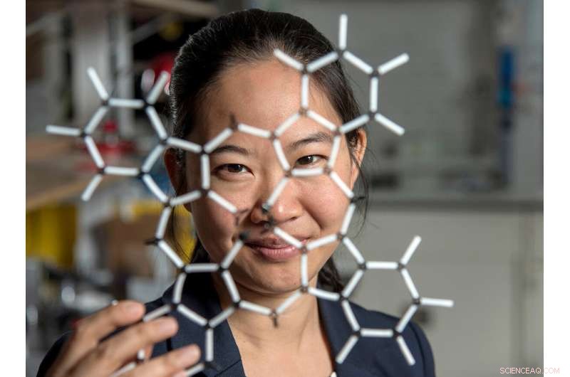
Pinshane Huang and her researchers discovered how to view structures in molecules, which opens a whole new realm of scientific possibilities. Credit:Heather Coit/The Grainger College of Engineering
How can you temper a molecule's environment? By using graphene.
Graphene, a single layer of carbon atoms that form a tight, hexagon-shaped honeycomb lattice, dissipates damaging reactions during imaging.
Stabilizing the small molecule's environment was only one issue the Illinois researchers had to manage. The team also had to limit its use of electrons, as low as one-millionth the number of electrons normally used, to illuminate the molecules.
Low doses of electrons ensure that the molecules are still moving enough for the researchers to capture an image.
"The way I like to think about it is the molecule doesn't like to be bombarded by higher-energy electrons, but we need to do that to be able to see the structure, and graphene helps dissipate some of that charge away from the molecule so that we can actually get a nice image of it," Janicek said.
Unfortunately, once captured, the molecules were nearly invisible in the image.
"When they take a low-dose image, it initially looks like noise or TV static—almost like nothing is there," Huang said.
The trick was to isolate the atomic structures from that noise by using a Fourier transform—a mathematical function that breaks down the small molecule's image—to see its spatial frequency.
"We took images of hundreds of thousands of molecules and added them together to build a single, clear image," Kharel said.
This averaging approach allowed the team to create crisp images of the molecules' atoms without damaging the integrity of any individual molecule.
"Month after month, week after week, our resolution improved," Huang said. "And then one day, my students came in and showed me the individual carbon atoms—that's a major achievement. And of course, it comes after all this deep knowledge that they have gained to design an imaging experiment and how to unlock data from what looks like nothing."
This collective discovery is paving the way for many more structural molecule imaging findings.
"There's been this whole field of small molecules that have been left out in the cold, so to speak. We're shining a light on how do we get there as a field? How do we make this thing that for us right now is so hard?" Huang said. "One day it won't be—that's the hope."
The Illinois researchers' efforts are the first big step in turning that dream into reality.
"One day, this will be the way we solve the structure of a small molecule," Huang said. "People will simply throw the molecule in the electron microscope, take a picture and be done."
That dream inspires Huang and her Illinois team to keep the course.
"That's potentially life-changing, and we've made it exist," Huang said. "We haven't yet made it easy, but imaging techniques like this will change so much of science and technology." ESR-STM sur les molécules simples et les structures à base de molécules